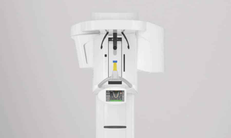
Digital volume tomography (DVT)
In dentistry, DVT stands for digital volume tomography. This is a three-dimensional imaging procedure that is used specifically for diagnostics in the head and jaw area.

CBCT provides precise 3D images of teeth, jaws and surrounding structures and is often used for
- Implant planning: To precisely measure the jawbone and nerve course and determine the optimal position for implants.
- Wisdom tooth removal: To assess the position of the wisdom teeth in relation to the surrounding nerves and structures.
- Orthodontic diagnostics: For the assessment of misaligned teeth or other orthodontic problems.
- Temporomandibular joint disorders: For the examination of problems in the temporomandibular joint.
CBCT is particularly advantageous because it provides a more detailed image compared to conventional X-ray images, while at the same time exposing the patient to less radiation than a computer tomography (CT).
In endodontics, i.e. the science of root canal treatment, digital volume tomography (DVT) is increasingly being used to obtain detailed three-dimensional images of teeth and their root canals.
Here are some specific applications and advantages of DVT in endodontics:
Applications of DVT in endodontics
1. recording the root canal anatomy: CBCT enables precise visualization of the root canals and their branches, which is crucial for diagnosis and treatment planning. It shows the number of root canals and their shape, including the branches and curvatures that are often difficult to recognize.
2. diagnosis of root canal problems: In complicated cases, such as necrotic or infected root canals, CBCT helps to identify the extent of the infection or the presence of abscesses and bone loss.
3. treatment planning: The detailed 3D images facilitate precise planning of root canal treatment by helping the dentist to determine the best course of action and minimize the risk of complications.
4. monitoring the success of treatment: After treatment, CBCT can be used to monitor the healing process, detect any remaining infection and check the integrity of the root filling.
5. visualization of microcracks and fractures: CBCT can also be used to identify microscopic cracks or fractures in the tooth that may not be visible by conventional radiographs.

Advantages of DVT
Detailed 3D imaging: CBCT provides a more comprehensive view of tooth and root canal anatomy compared to traditional 2D x-rays, resulting in a more accurate diagnosis.
Reduced radiation exposure: Compared to conventional computed tomography (CT), CBCT generally has a lower radiation exposure for the patient.
Improved diagnostics: Detailed imaging allows problems to be detected earlier and more precisely, which improves treatment outcomes.
Overall, CBCT in endodontics helps to refine diagnosis and optimize treatment, improving patient outcomes.
OPENING HOURS
Mon – Thu: 8 a.m. – 7 p.m.
Fri: 8 a.m. – 2 p.m.
and by appointment
Emergency dental service
Tel: +49 30 89004333